
A new study published in Nature has uncovered a universal map of the tumor microenvironment shared across 17 different cancer types—and, crucially, demonstrated that its features can be detected from a blood test to predict whether patients will respond to immunotherapy.
The work, led by Aaron Newman, PhD, at Stanford University and Aadel Chaudhuri, MD, PhD, of the Mayo Clinic describes nine distinct “spatial ecotypes”—stereotyped communities of immune and structural cells that organize themselves in consistent patterns around tumors, regardless of the cancer type. The researchers then developed an AI-based algorithm capable of inferring the proportions of those ecotypes from circulating tumor DNA in plasma, translating a tissue-level biological map into a minimally invasive liquid biopsy readout.
A universal architecture
The researchers studied more than 100 tumor specimens across 10 cancer types, using their tools to map gene expression patterns across nine cell types at varying locations throughout the tumor. They identified nine distinct spatial ecotypes—neighborhoods roughly the diameter of a human hair—and found that these patterns were conserved across all tumors studied. Some ecotypes clustered at the border between tumor and healthy tissue; others appeared deeper within the tumor mass. Several of the ecotypes correlated with immunotherapy response, pointing to potential clinical utility in guiding treatment decisions.
The central discovery is that tumors are not randomly organized. Across carcinomas of the breast and prostate, melanoma, and more than a dozen other cancer types, the same nine spatial ecosystems appear—each with a distinct cellular composition, gene expression signature, and physical location relative to the tumor mass.
“Cells exist not in isolation, but in little communities or neighborhoods or social networks,” said Newman. “These characteristics were highly stereotypical, highly conserved across different carcinomas and melanomas. Each spatial ecosystem has its own common features that differ from other spatial ecosystems—you can think of them as the parts list that most tumors are made of when we think about the soil that nurtures the cancer cells.” And, each spatial ecotype has its own internal social network—the cellular behaviors, or genetic programs it is carrying out, are influenced by the cells around it.
Chaudhuri, a clinician scientist, offered a grounding analogy. “Whether you’re in Tokyo or Minneapolis or San Francisco, you have a downtown, and as you go farther out, you have Midtown, suburbs, and then rural farmland. That’s how it is for the tumor microenvironment as well—close to the tumor cells, you have spatial ecotypes seven, eight, and nine; go farther away and you have spatial ecotypes one, two, three, and four. That map is consistent across all these different cancer types.”
Predicting immunotherapy response
The clinical implications center on immune checkpoint inhibitors, a class of drugs that has transformed oncology but still fails a significant portion of patients. Analyzing 15 immunotherapy cohorts in tissue, the team identified two spatial ecotypes—SE7 and SE8—characterized by T cells positioned at and within the tumor, associated with a pro-inflammatory, anti-tumor immune response, and strongly linked to immunotherapy benefit. Conversely, a fourth ecotype, SE4, associated with wound-healing biology, was strongly predictive of resistance.
“Higher levels of SE7 and SE8 strongly forecast the patient’s response to immune checkpoint inhibitors,” said Newman. “And conversely, higher levels of SE4 portend a worse outcome—resistance to immune checkpoint inhibitors.”
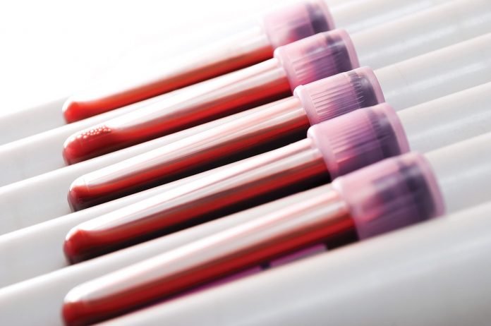
Those tissue-based findings were then validated in blood. Using a deconvolution algorithm the Newman lab developed—the same class of computational approach used to identify cell-type proportions from bulk sequencing data—the team showed that plasma samples taken before treatment could recapitulate the spatial ecotype signals found in paired tumor biopsies, and that those liquid-biopsy-derived ecotype levels predicted patient outcomes with striking fidelity.
“Without looking at their tumor biopsy, just taking a blood sample, we could determine the levels of the liquid spatial ecotypes from their tumors directly from the blood,” Newman said. “Those levels very strongly predicted their outcomes once they received immunotherapy.”
Beyond the single biopsy
Perhaps the most consequential aspect of the platform is what it enables over time. Today, a patient’s tumor microenvironment can be assessed only at the moment of biopsy—a single, static snapshot that may be compromised by sampling bias and cannot be repeated without invasive procedures.
“There is no existing clinical assay to access these features of the cancer over time,” Newman noted. “For the first time, this gives us those insights.”
The team has already begun longitudinal work, with funded studies planned to track melanoma patients serially through immunotherapy—monitoring how spatial ecotype levels shift on treatment and whether those dynamics outperform existing tools like CT imaging, which can struggle to distinguish true progression from pseudo-progression.
Chaudhuri emphasized the broader paradigm shift. “For years and years, we have been completely tunnel-visioned on malignant tumor cells and their mutations, measuring their mutations as surrogates for MRD and things like that,” he said. “For the first time ever, we’ve opened our eyes and we can look at the tumor microenvironment, which is absolutely critical for precision oncology.”
From lab to clinic
The technology has been licensed to a startup company, Liquid Cell Dx, co-founded by Newman and Chaudhuri, with the goal of developing and validating a clinical-grade assay. The team has already presented blinded clinical validation data at AACR, applying cut points learned from the Nature paper cohort to an entirely independent patient population at a separate institution—without knowledge of outcomes until unblinding.
Active translational programs are now underway in non-small cell lung cancer, muscle-invasive bladder cancer, and mesothelioma, examining spatial ecotypes in the context of chemotherapy combinations, neoadjuvant settings, and radiotherapy.
Newman was recently awarded an AACR Trailblazer Award for the serial monitoring work in melanoma.
“The goal here is for this not to just end at the Nature paper, for it to benefit humanity,” said Chaudhuri. “We want to take the technology, get it into patients, develop it into a clinical assay, and really show that we can bring next-level precision into oncology.”